Karyotype Definition, Test, Functions, and Human Chromosome Numbering – Karyotype (derived from the Greek word: karyon = for “node” and type = for “shape”), symbolizes a collection of diploid chromosomes (2N) of the somatic cells of an organism.
By understanding what a karyotype is, it becomes easier to understand the importance of identifying numerical and structural chromosome changes.
Karyotypes can be represented through chromosome images (karyograms) or by the size of chromosomes in photographic schemes (idiograms).
By arranging a karyotype, it is possible to determine normality or abnormalities (chromosomal syndrome), caused by mutagenic changes, polysomy or monosomy.
In the human species, somatic cells have 46 chromosomes (2N = 46), grouped into 23 pairs, namely: 22 pairs of autosomal and 01 pairs of se**xual allosome, which distinguishes the se**x of an organism in both males and females.
- Normal male karyotype → 46, XY
- Karyotype of normal women → 46, XX
Read also:
Homologous Chromosomes: Definition, Etymology, Pair, Function, and Characteristics
Karyotype Definition
A karyotype is a collection of chromosomes that a person has. The term also refers to laboratory techniques that produce images of such chromosomes. Karyotypes are used to look for abnormal numbers or chromosomal structures.
A karyotype is a test to identify and assess the size, shape and number of chromosomes in a sample of body cells. Additional or missing chromosomes, or abnormal positioning of parts of chromosomes, can cause problems in the growth, development, and functioning of a person’s body.
While pregnant, the doctor can ask the patient to do some prenatal examinations to look for certain genetic abnormalities and chromosomes. This procedure may be requested as the pregnancy enters the first trimester and then continues in the second trimester.
If the patient gets results that are within the normal range, then the patient does not require further tests. But if there are indications that show signs of abnormality, the doctor will offer a follow-up test so that the patient can know for sure if the baby being conceived has a genetic or chromosomal problem. Doctors will need a small sample of the baby’s cells to examine his chromosomes.
When to do karyotype test?
This test may be performed:
- In couples who have a history of miscarriage
- To check for children or infants with unusual features or late development
Bone marrow or blood tests could be performed to identify the Philadelphia chromosome, which is found in 85% of people with chronic myelogenous leukemia (CML). Amniotic fluid tests are carried out to check developing babies for chromosomal problems.
Karyotype Function
Karyotype is done for:
- Determines whether the older persons’ chromosome has abnormalities that can be passed on to the child;
- Determine chromosomal defects to prevent women from causing miscarriage;
- Determines whether chromosomal defects are present in the fetus. This examination can also be done to determine if chromosomal problems may have caused the fetus to fail;
- Determine the cause of the baby’s birth defect;
- Helps determine the right treatment for some types of cancer;
- Identify a person’s gender by determining the presence of the Y chromosome.
Karyotype testing can be done using almost any cell or tissue from the body. A karyotype test is usually performed on a blood sample taken from a vein. For testing during pregnancy, it may also be done on samples of amniotic fluid or the placenta.
Human chromosome numbering
Each chromosome has been numbered based on its size. The largest chromosome is chromosome 1. Therefore chromosome 18 is one of the smallest chromosomes in humans.
The basic number of chromosomes in the somatic (body) cells of an individual or species is called a somatic number and is set at 2n. Thus, in humans 2n = 46. In genital cells, the number of chromosomes is N (human: n = 23).
Thus, in normal diploid organisms, chromosomes are present in two copies. There may be, or may not, be genital chromosomes. Polyploid cells have multiple copies of chromosomes and haploid cells have a single copy. The study of a whole set of chromosomes is sometimes known as karyology.
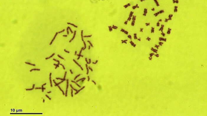
Chromosomes are described (by reorganizing microphotographs) in a standard format known as a karyogram or idiogram: paired, arranged by size and position of the flashlight for chromosomes of the same size.
It can be used for many purposes; like, to indicate the genetic disease, the se**x of the animal and the amount of its diploid. In humans, cytologists have used a karyotype to help identify genetic abnormalities a baby may have before he was born.
Karyotypes can be studied to gather information about past evolutionary events, such as polyploidy.
Conclusion
The karyotype definition is the number and appearance of chromosomes in the nucleus of eukaryotic cells. The term is also used for a complete set of chromosomes in a species, or individual organism.
Karyotype describes the number of chromosomes, and what they look like under a light microscope. Attention is paid to the length, position of the flashlight, the difference between the genital chromosomes, and other physical characteristics. The preparation and study of karyotype is part of Cytogenetics (cytology and genetics).
Source:
- Image: Doc. RNDr. Josef Reischig, CSc., CC BY-SA 3.0 https://creativecommons.org/licenses/by-sa/3.0, via Wikimedia Commons.
- Video: Nicole Lantz


 Skincare Solutions & Skin Health
 Mental Health, Stress & Emotional Wellbeing
 Sleep, Energy & Daily Performance